The world famous 3D viewer
The dentist reconstructs the patient's DICOM file generated through cone beam CT imaging in
three dimensions to help the user accurately analyze and read the CT results.
You can display three viewer modes on one screen, from MPR, Panorama, and Implant. It is advantageous for patient counseling because comparison and contrast are possible at a glance.
It can develop its own engine and organize images faster.
Various edits of reconstructed 3D images can be made according to user convenience (zoning, cropping, length measurement, coordinate acquisition, etc.)
Users can adjust the roughness of the image in real time and check it carefully as needed.
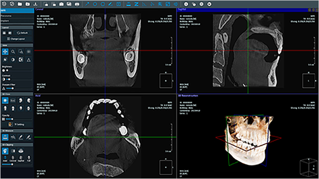
MPR
The reconstructed 3D image screen and three 2D images, axial, coronal, and sagittal, are customized at a glance. Various configurations can be edited.
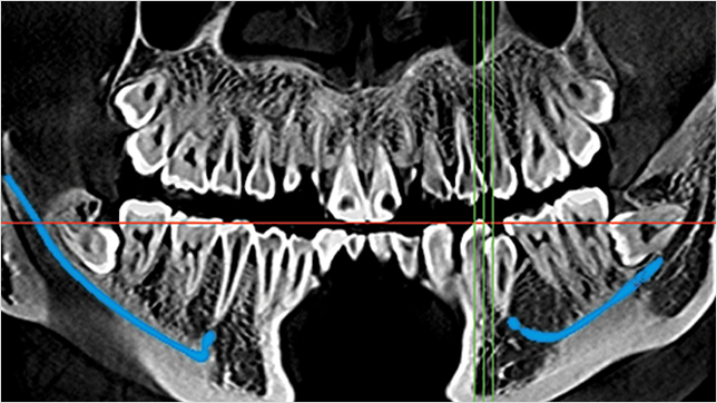
Panorama
Configured to facilitate comparison/contrast between panorama and axial screens through cross-section algorithms. Canal Drawing function to locate and draw neural tubes for treatment
Implant
Establish implantation simulation, bone density, and neural tube position of implant actually used in the treatment location to notify the success of the procedure.
MPR
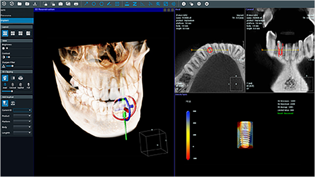
Implant
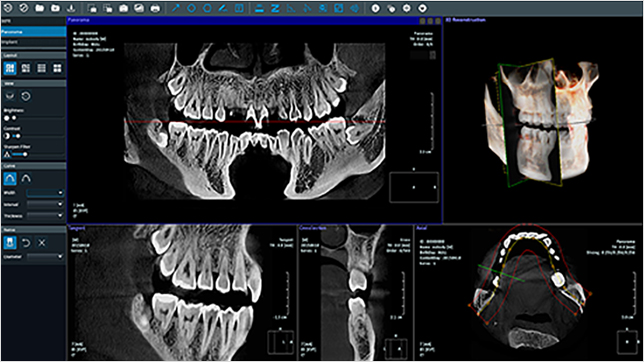
Panorama
Real-time
The user arbitrarily adjusts the degree of roughness and corrects the screen in real time.
Delicate
Adjustable sharpness according to delicate roughness differences.
Communication
Provide visual information during prior consultation with the patient to help consent to the procedure.
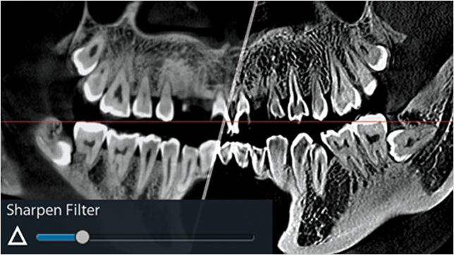
Real Sharpen
User control
Guess the location of the neural tube that does not appear on the cone-beam CT scan, and display it directly by the user.
Cross section
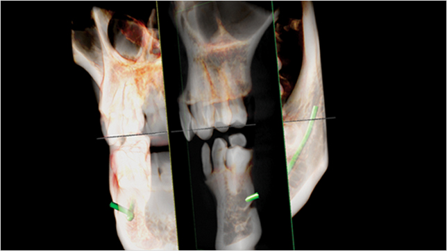
The position of the neural tube drawn on Panorama is automatically applied to 3D images and cross-section images.
Intuitive explanation
Clearly distinguished from CT images, allowing intuitive explanation to patients.
Cross Section
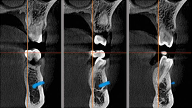
Cross Section
Cross Section
Address 1006, 43, Digital-ro 34-gil, Guro-gu, Seoul, Republic of Korea
Tel +82-2-6956-1465
Fax +82-2-6949-0363
Mail sales@sugarpole.com