the next generation of medical imaging. Explore, analyze, and understand medical data in stunning 3D. Built for precision and efficiency, Sugar 3D transforms the way you interact with CT, MR, and other DICOM images, empowering more accurate diagnoses and improved patient care.
![]()
Fast and precise 3D reconstruction for efficient diagnostics.
![]()
View lesions in three dimensions from multiple perspectives.
![]()
Intuitive interface that's easy for non-technical users to use.
![]()
Support for advanced tools for precise visualization.
![]()
Advanced 3D analysis tools for precise clinical evaluation.
![]()
Scalable configurations to take advantage of additional advanced features.
CT DICOM data is rendered in real time using a GPU-based engine, enabling smooth and delay-free 3D rendering. 3D volumes are generated instantly without conversion, allowing seamless interaction such as rotate, zoom, and slice.
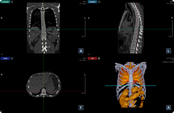
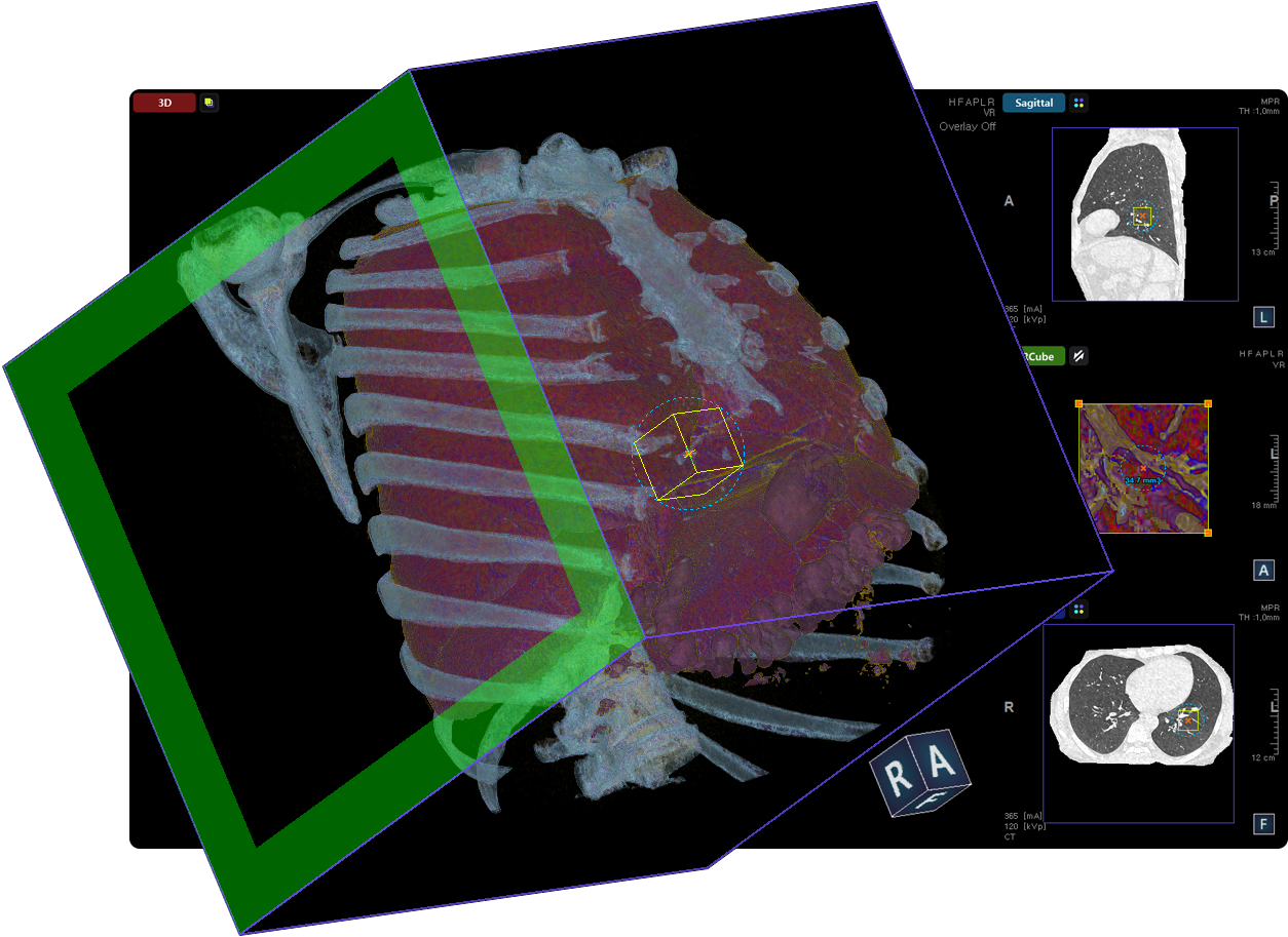
Axial, Coronal, Sagittal and 3D View
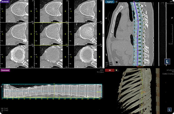
Panoramic and Sectional View
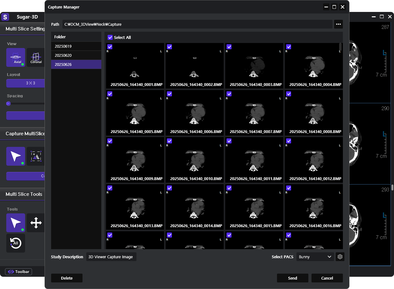
Multi Slice View
3D Multi Rotate View
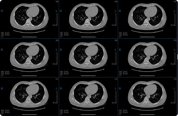
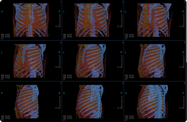
Standard planes such as Axial, Coronal, and Sagittal offer comprehensive anatomical visualization. Panoramic and sectional views provide extended visibility along curved or complex regions. Multislice mode enables side-by-side comparison of sequential slices, ideal for tracking lesion location and extent.
The intuitive user interface and quick-access tools minimize the learning curve and streamline diagnostic workflows. Customizable layouts and keyboard shortcuts enhance user efficiency and adaptability across different use cases.
Axial, Coronal, Sagittal and 3D View
Panoramic and Sectional View
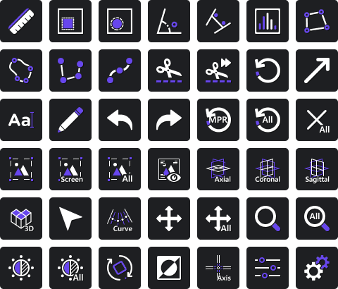
Built-in tools for measuring distance, angle, area, and volume are complemented by features like clipping, segmentation, and color mapping. These capabilities support precise anatomical analysis and pre-surgical planning with high visual clarity.
VOI lets you highlight specific regions for focused analysis, while the VR Cube enables intuitive 3D navigation and multi-angle exploration.
Comes with a comprehensive library of implants, allowing precise control of position, angulation, and depth in a 3D environment. Supports accurate pre-surgical planning through interactive simulation, and serves as a powerful tool for patient communication and case presentation.

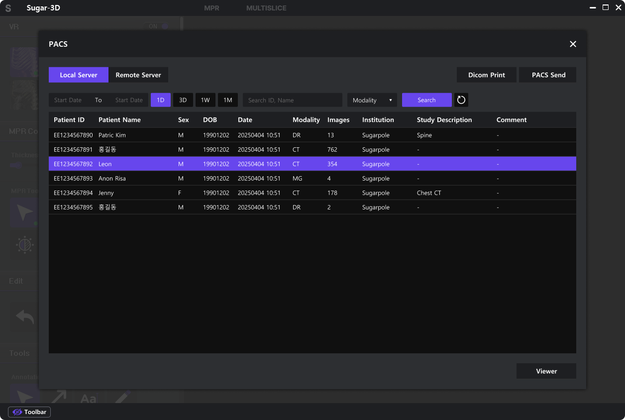
Seamlessly integrates with hospital PACS systems, allowing direct use of original DICOM data without conversion. Ensures consistency and accuracy diagnostic imaging.
Captured images can be saved in DICOM format and quickly shared across departments or systems. Ideal for clinical collaboration and patient reporting.
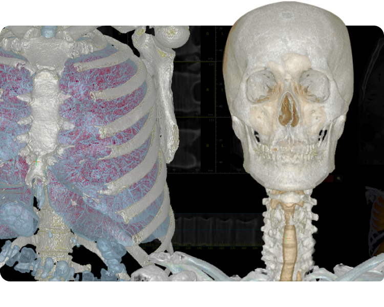
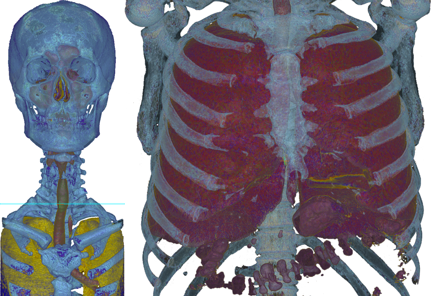
Provides precise 3D visualization of thoracic anatomy including airways, vessels, and lymph nodes, allowing accurate localization and measurement of lesions. Supports surgical planning by identifying tumor margins and critical structures to avoid. Ideal for complex procedures requiring in-depth anatomical insight through multiple viewing modes.

Offers comprehensive 3D visualization of bone structures and joint alignments, enabling accurate diagnosis of fractures and assessment of surrounding tissues. Simulates implant positioning and orientation to support precise surgical planning. Also facilitates postoperative evaluation through detailed cross-sectional comparisons.
Combines axial slices with 3D volume rendering to enable multidimensional analysis of complex lesions and anatomical abnormalities. Visualizes density variations, lesion margins, and relationships with surrounding structures, enhancing both diagnostic confidence and reading speed. Captured images can also be instantly shared via PACS, facilitating fast and accurate communication for multidisciplinary collaboration.
| Function | Standard | Premium |
|---|---|---|
| 3D Volume Rendering | ✔ | ✔ |
| Multiple View Modes | ✔ | ✔ |
| Advanced Tools | ✔ | ✔ |
| Expert-Level Tools | ✔ | ✔ |
| Implant Simulation | ✔ | |
| PACS Integration | ✔ | |
| Dicom Function | ✔ |
| Category | Minimum | Recommended |
|---|---|---|
| OS | Microsoft Windows 10 | Microsoft Windows 10, 11 |
| CPU | 1.5GHz+ | 3GHz Dual Core+ |
| RAM | 8GB | 16GB+ |
| GPU | NVIDIA/AMD Graphics Card | NVIDIA/AMD Graphics Card(4GB+) |
| Display Resolution | 1280X1024 | 1920X1080 |
| Storage | 200MB HDD install space | Fast SSD(Image Data Storage) |
Address #1111, 306, Digital-ro, Guro-gu, Seoul, Republic of Korea
Tel +82-2-6956-1465
Fax +82-2-6949-0363
Mail sales@sugarpole.com